Beranda
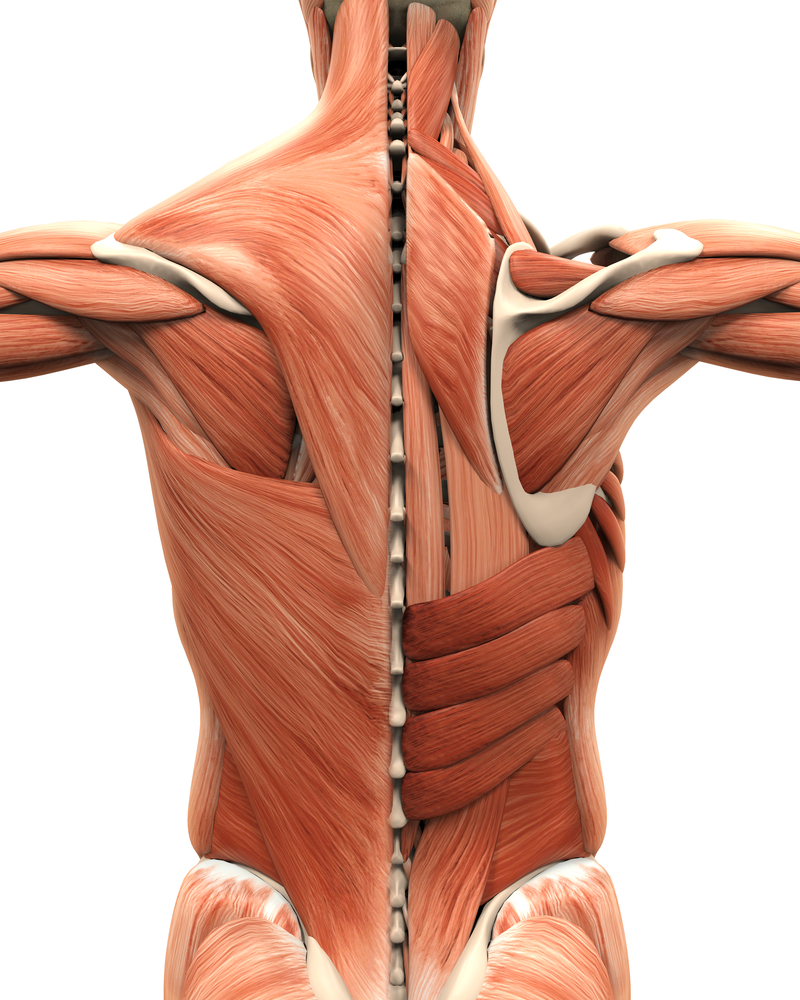
/ Back Muscles Anatomy : Quadratus Lumborum — EMPA / They provide movements of the spine , stability to the trunk, as well as the coordination between the movements of the limbs and trunk.
Back Muscles Anatomy : Quadratus Lumborum — EMPA / They provide movements of the spine , stability to the trunk, as well as the coordination between the movements of the limbs and trunk.
Insurance Gas/Electricity Loans Mortgage Attorney Lawyer Donate Conference Call Degree Credit Treatment Software Classes Recovery Trading Rehab Hosting Transfer Cord Blood Claim compensation mesothelioma mesothelioma attorney Houston car accident lawyer moreno valley can you sue a doctor for wrong diagnosis doctorate in security top online doctoral programs in business educational leadership doctoral programs online car accident doctor atlanta car accident doctor atlanta accident attorney rancho Cucamonga truck accident attorney san Antonio ONLINE BUSINESS DEGREE PROGRAMS ACCREDITED online accredited psychology degree masters degree in human resources online public administration masters degree online bitcoin merchant account bitcoin merchant services compare car insurance auto insurance troy mi seo explanation digital marketing degree floridaseo company fitness showrooms stamfordct how to work more efficiently seowordpress tips meaning of seo what is an seo what does an seo do what seo stands for best seotips google seo advice seo steps, The secure cloud-based platform for smart service delivery. Safelink is used by legal, professional and financial services to protect sensitive information, accelerate business processes and increase productivity. Use Safelink to collaborate securely with clients, colleagues and external parties. Safelink has a menu of workspace types with advanced features for dispute resolution, running deals and customised client portal creation. All data is encrypted (at rest and in transit and you retain your own encryption keys. Our titan security framework ensures your data is secure and you even have the option to choose your own data location from Channel Islands, London (UK), Dublin (EU), Australia.
Back Muscles Anatomy : Quadratus Lumborum — EMPA / They provide movements of the spine , stability to the trunk, as well as the coordination between the movements of the limbs and trunk.. Bodybuilder showing his back and biceps muscles, personal fitness trainer. Back muscles, functions and exercises: These sections are cervical (neck), thoracic (upper and middle back), lumbar (lower back), and sacrum (tailbone). Muscle anatomy gluteus 12 photos of the muscle anatomy gluteus gluteus muscle anatomy ct, gluteus muscle anatomy mri, human muscle anatomy gluteus maximus, muscle anatomy gluteus, muscle anatomy of gluteal, human muscles, gluteus muscle anatomy ct, gluteus muscle anatomy mri, human muscle anatomy. Anterior rami of spinal nerve innervate them.
(2017, elsevier) should be consulted. Memorize all the muscle facts with the help of muscle cheat sheets. Anatomy of the back muscles the latissimus dorsi muscles (also known as the lats) are the largest muscles of the back. The surface muscles of the upper back include the trapezius muscles (traps) and posterior deltoids. Understanding lower back anatomy is key to understanding the root of lower back and hip pain.
Muscle and ligament pain in the lower back from buxtonosteopathy.co.uk By far the most common cause of back pain is muscle strain. The muscles of the back muscles make up a large part of the anatomy (structure) of the back. Leaning back to straight vertical and all points in between. The back muscles are divided into two large groups: Your lower back (lumbar spine) is the anatomic region between your lowest rib and the upper part of the buttock. These muscles include the large paired muscles in the lower back, called erector spinae, which help hold up the spine, and gluteal muscles. This blog post article is an overview of the muscles of the lumbar spine of the trunk. Muscle anatomy gluteus 12 photos of the muscle anatomy gluteus gluteus muscle anatomy ct, gluteus muscle anatomy mri, human muscle anatomy gluteus maximus, muscle anatomy gluteus, muscle anatomy of gluteal, human muscles, gluteus muscle anatomy ct, gluteus muscle anatomy mri, human muscle anatomy.
Balance the weight of your head on top of your spine evenly distribute weights from your upper body into the lower extremities
These structures work together to support the body, enable a range of movements, and send messages from the brain to the. Your lower back (lumbar spine) is the anatomic region between your lowest rib and the upper part of the buttock. The deep muscles develop in the back called intrinsic muscles. Back pain is the second most common type of pain in adults (the most common being headaches). The surface muscles of the upper back include the trapezius muscles (traps) and posterior deltoids. The back comprises interconnecting nerves, bones, muscles, ligaments, and tendons, all of which can be a source of pain. The human spine is composed of 4 sections of vertebrae. The multifidus, a long muscle that travels nearly the entire length of the back.it helps to stabilize and rotate the lower back, and additionally takes some. They start at the top of the neck and go down to the tailbone. Leaning back to straight vertical and all points in between. Anatomy chart courtesy of fcit the latissimus dorsi muscles (also known as the lats) are the largest muscles of the back. Bodybuilder showing his back and biceps muscles, personal fitness trainer. This is an online quiz called back muscles there is a printable worksheet available for download here so you can take the quiz with pen and paper.
On this page, you'll learn about each of these muscles, their locations and functional anatomy. These muscles include the large paired muscles in the lower back, called erector spinae, which help hold up the spine, and gluteal muscles. (2017, elsevier) should be consulted. The intrinsic back muscles are found deeper to the extrinsic muscles, separated from them by the thoracolumbar fascia. Your lower back (lumbar spine) is the anatomic region between your lowest rib and the upper part of the buttock.
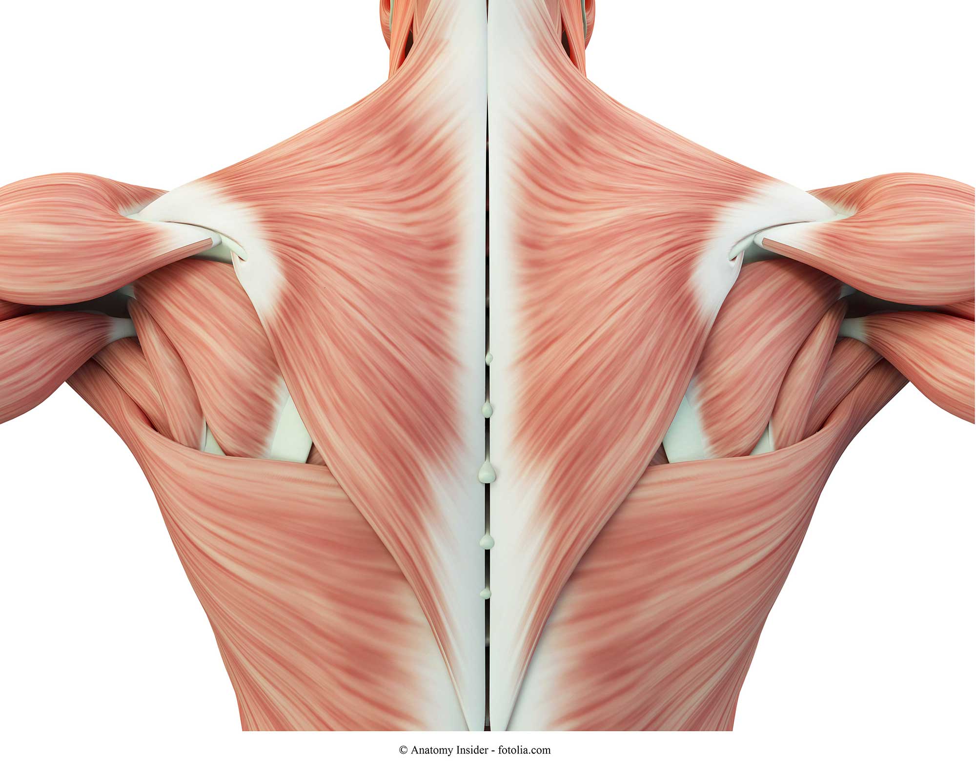
Muscles Move and Support the Spine from www.spineuniverse.com All these muscles are therefore associated with movements of the upper limb. Back muscles, functions and exercises: Back pain is common and might be caused by a problem with a muscle. Balance the weight of your head on top of your spine evenly distribute weights from your upper body into the lower extremities Back muscles the muscles of the back are a group of strong, paired muscles that lie on the posterior aspect of the trunk. Muscles of the back can be divided into superficial, intermediate, and deep group. These muscles determine body posture and also regulate the three basic movements of the trunk: These muscles give height and breadth to back development.
They start at the top of the neck and go down to the tailbone.
Back muscles the muscles of the back are a group of strong, paired muscles that lie on the posterior aspect of the trunk. The back muscles are anatomically layered into superficial (extrinsic) and deep (intrinsic) muscles. Muscles of the lumbar spine. Muscle anatomy gluteus 12 photos of the muscle anatomy gluteus gluteus muscle anatomy ct, gluteus muscle anatomy mri, human muscle anatomy gluteus maximus, muscle anatomy gluteus, muscle anatomy of gluteal, human muscles, gluteus muscle anatomy ct, gluteus muscle anatomy mri, human muscle anatomy. They provide movements of the spine , stability to the trunk, as well as the coordination between the movements of the limbs and trunk. Related posts of muscles of the lower back and buttocks diagram muscle anatomy gluteus. Three types of back muscles that help the spine function are extensors, flexors and obliques. Back muscles, functions and exercises: Memorize all the muscle facts with the help of muscle cheat sheets. (2017, elsevier) should be consulted. Anatomy chart courtesy of fcit the latissimus dorsi muscles (also known as the lats) are the largest muscles of the back. They start at the top of the neck and go down to the tailbone. Superficial back muscles, intermediate back muscles and intrinsic back muscles.the intrinsic muscles are named as such because their embryological development begins in the back, oppose to the superficial and intermediate back muscles which develop elsewhere and are therefore classed as extrinsic muscles.
The back consists of the spine, spinal cord, muscles, ligaments, and nerves. The human spine is composed of 4 sections of vertebrae. Anterior rami of spinal nerve innervate them. Tutorials on the anatomy and actions of the back muscles, using interactive animations, diagrams, and illustrations. Back pain is common and might be caused by a problem with a muscle.
Trapezio, Pettorali, Dorsali e Muscoli della Spalla from www.fisioterapiarubiera.com Back muscles the muscles of the back are a group of strong, paired muscles that lie on the posterior aspect of the trunk. The extensor muscles are attached to back of the spine and enable standing and lifting objects. Anterior rami of spinal nerve innervate them. Back pain is the second most common type of pain in adults (the most common being headaches). Leaning back to straight vertical and all points in between. The back muscles are anatomically layered into superficial (extrinsic) and deep (intrinsic) muscles. Three types of back muscles that help the spine function are extensors, flexors and obliques. Balance the weight of your head on top of your spine evenly distribute weights from your upper body into the lower extremities
Leaning back to straight vertical and all points in between.
Leaning back to straight vertical and all points in between. The superficial back muscles are situated underneath the skin and superficial fascia. Superficial back muscles, intermediate back muscles and intrinsic back muscles.the intrinsic muscles are named as such because their embryological development begins in the back, oppose to the superficial and intermediate back muscles which develop elsewhere and are therefore classed as extrinsic muscles. Back pain is common and might be caused by a problem with a muscle. This curve, called lordosis, helps to: Strong man flexing his muscles. The extrinsic back muscles are located in the back, but act to produce movements of the shoulder and assist respiration. Muscles of the lumbar spine. Anatomy chart courtesy of fcit the latissimus dorsi muscles (also known as the lats) are the largest muscles of the back. By far the most common cause of back pain is muscle strain. They provide movements of the spine , stability to the trunk, as well as the coordination between the movements of the limbs and trunk. Anatomy of back muscles your back consists of three distinct layers of muscles, namely the superficial layer, the intermediate layer, and the deep layer. The back muscles are anatomically layered into superficial (extrinsic) and deep (intrinsic) muscles.